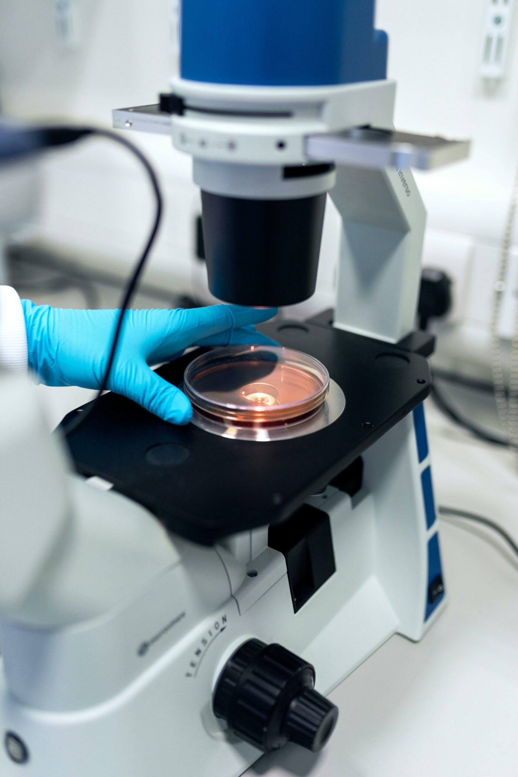
In vitro methods use tissues, micro-organisms, cells or other biological materials. The most promising of these are based on classic cell culture techniques, to produce inexpensive and rapid 2 and even 3-dimensional models for the analysis of various organs and tissues. Results in human medicine are all the more reliable when using cells and tissues of human origin.
Examples of “in vitro” methods?
Stem cells of human origin
There are several types: tissue-specific stem cells, umbilical cord stem cells, pluripotent stem cells, including embryonic stem cells and induced pluripotent stem cells (iPS cells). The latter are mature cells which, after “reprogramming” (de-differentiation then re-differentiation into the desired cell type), can give rise to all the body’s cell types, whether normal or pathological (if they come from a patient suffering from a given disease).
The hopes pinned on these techniques cover a wide range of fields, including toxicology, cell therapy and regenerative medicine; they are also invaluable tools in fundamental research. Biobanks of pluripotent stem cell lines are being set up.
Organoids
They are made from cell cultures (tissue precursor cells, stem cells), aiming to present human organ functionalities in 3 dimensions. The technique has been developed since 2013. They are increasingly used in both healthy and pathological tissues, as cellular models of human diseases. Their applications are manifold: regenerative medicine, toxicity testing, drug efficacy testing, infection modeling, personalized medicine… and this for virtually every organ, including the brain or retina.
These organoids are not capable – in the current state of research – of reproducing all biological responses like a real organ, but they do enable researchers to study a variety of physiological responses to specific manipulations and treatments. They also make it possible, for a given patient, to test the efficacy of a potential drug using his or her own cells.
Organs on a chip
They have been developed thanks to advances in both nanotechnology and cell culture. Thanks to microfluidic systems (microchannels), these devices can reproduce the structure and function of an organ (or several organs): skin, lung, liver, heart muscle, bone, kidney, intestine, brain…
These organs-on-a-chip are made of a transparent, flexible polymer and contain hollow microfluidic channels lined with living human cells (from a biopsy, or stem cells, or immortalized cell lines). Microfluidic channels contain tiny quantities of liquid ranging from less than a micron to a few millimeters. They are equipped with mechanical forces that can mimic the physical environment of organs, such as respiratory movements (lung-on-a-chip) or peristaltic-type movements (intestine-on-a-chip); a micro-perfusion system can simulate the blood circuit. When nutrients, air, blood or drugs are added, the cells reproduce some of the organ’s key functions. Such chips can include several interconnected organs (multi-organ chips).
These techniques can be used for regulatory efficacy and toxicity studies, personalized medicine, and pathology studies (using tumor cells, for example).
The players are mainly start-ups marketing prototypes. In fact, it’s a fast-growing industry.
Bioprinting
Tissue printing is a tissue engineering and regenerative medicine technology that involves researchers from different disciplines: medicine, engineering, biology and chemistry.
There are various 3D printing technologies. The general principle is to use computer-controlled processes to join materials or liquids together, layer by layer, to create three-dimensional objects, in this case, artificial biological tissue. 3D bioprinting uses cells and other biological products to build living tissue. Researchers therefore had to find materials and printing processes compatible with living cells and tissues. They also had to find suitable materials to build scaffolds that contain and shape the biomaterials into the desired form. These scaffolding materials can be natural or synthetic. Several methodological approaches have been developed (laser, “drop” deposition, micro-extrusion…).
Potential applications are vast: in vitro models of pathologies, drug testing (efficacy, toxicity), screening, regenerative medicine (for organ transplants in particular).
Certain technical obstacles remain. There is a trade-off between resolution, compatibility with cell deposition, cell viability and mechanical stability. Rapid progress is expected, however.
Bioprinting can be combined with organ-on-a-chip technology to study physiological functions.
High-throughput screening
It represents an automated method for testing the biological activities of thousands of chemicals that were previously tested on animals; the idea is to have a large number of different molecules react simultaneously with a given substrate. This qualitative leap in procedure drug discovery has been achieved thanks to a synergy between chemistry, biology, engineering and computer science. In the USA, this technique is supported by a government initiative called “Toxicology in the 21st Century” (Tox21), to “develop better toxicity assessment methods to quickly and efficiently test whether certain chemical compounds have the potential to disrupt processes in the human body that can lead to adverse health effects”. The aim is to provide predictive models of biological response in vivo.
In Europe, Eurotox (federation of European toxicologists and European toxicology societies) pursues similar objectives.
High-throughput testing is also used with organ-on-a-chip. This technology is currently still too expensive for widespread use, but work is underway to make it more cost-competitive so that it can replace conventional models.


